Wednesday, February 15, 2012

Saving the Brain

I want to revisit the topic of neurogenesis (the growth of new neuron cells) because while it may be that adult brains can form new neurons, they can't do so to the degree found with brains experiencing the critical period.  The critical period occurs when between late embryonic stages and early life.  This is when massive amounts of new neurons are created and preliminary synapses are growing towards as many targets as possible.
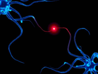
The power of a single synapse is weak, but together in a
circuit their power is incredible. 

After the critical period, there is a massive reduction in the number of connections between neurons.  The ones that remain are typically efficient enough to handle all the mental processes but the ability to "rewire" after this period is severely reduced.

However, some recent research is leading us to believe that there may be a way to reopen the critical period  and at least partially rewire our brains.  In fact, some antidepressants seem to be doing just that.  There's now evidence that anti-depressants like fluoxetine work by increasing BDNF (brain derived neurotrophic factor) which is hallmark of the critical period.  Fluoxetine requires chronic usage to get these effects but perhaps the antidepressant allows for the creation of new synapses and connections.  Maybe the rewiring is changing how information is processed in the emotional centers of the brain, thus relieving the depression of the patient.
A pill that changes how your brain is wired-
scary or amazing?

There's still plenty of research and trails we have yet to undergo before we understand all the mechanisms.  But this offers hope that neuroplasticity can be raised to the high level of malleability found during the critical period.

Tuesday, February 14, 2012

Roses are red, Violets are blue, Neurotransmitters make me love you!

In honor of today's Hallmark-funded holiday which is customarily filled with bright pink hearts and chalky candy handed out to all grade-schoolers, I decided to write about what makes us feel "in love."  Yes, you can read all the romance novels you want (*cough*Twilight*cough*) which expound on his chocolate eyes or her sensuous curves.  But when we move back to the basics, it's really just a soup of neurochemicals stirring around in your brain that cause that spark of love in your life.
Who actually eats these?

And truth be told, the biochemistry of attraction and love is still under research.  However we can safely say that there are 3 key neurotransmitters that make Valentine's Day special for all those people out there with their significant others.

The first and likely most recognizable is dopamine.  It plays many roles in the chemical signaling of the brain but is primarily associated with the reward pathway.  Whenever you get that rush of happiness from the first bite of chocolate cake, that's the dopamine kicking in.  In fact, addiction and the need to up the dosage of drugs over time like cocaine comes from the brain desensitizing due to the over use of dopamine.  And in the romantic sense, dopamine is the "drug" part of love.  It's the passionate, euphoric love that makes you believe there's nothing better than being with your partner.
How many roses do you think are sold each
 valentine's day?

And yet that's not quite enough.  Can you remember the first time you asked that girl to dance at the winter formal?  Or how about the anticipation before your first kiss?  Odds are your heart was pounding like a jack-hammer.  Norepinephrine is the neurotransmitter that provides this autonomic response.  It focuses your attention and physically primes you with those classic signs of nervousness.  Next time your hands turn clammy when you reach out towards your love interest, you can blame norepinephrine.

But these two neurochemicals play havoc on your brain if they're constantly swamping the circuits.  To keep an even keel yet stay connected to your significant other, we need oxytocin.  This compound is a long lasting messenger which doesn't give the same high as the others, but leads to a feeling contentment. Sometimes called the "cuddling neurotransmitter," oxytocin provides us with the companionship aspect of romantic relationships.  Without it we wouldn't have the same peace and stability in our long-term relationships.

Ok, so there are actually many more circuits and neurochemicals involved in love. It's an emotion not easily reduced to formulas and equations.  In my opinion, we could lecture on the biological components involved all day, but honestly I prefer to just listen to some Maroon 5 and eat chocolate covered strawberries!


If you want to hear more on this subject, I suggest checking out the original source:
http://www.radiolab.org/blogs/radiolab-blogland/2007/aug/28/this-is-your-brain-on-love/
Radiolab is a non-profit public radio program hosted by WNYC.  If you'd like to donate to their phenomenal program, please go to: https://pledge3.wnyc.org/epledge/desktop/radiolab/

Friday, February 10, 2012

Adult Neurogenesis- The Brain That Grows

When you first start to learn science in grade school, everything seems so set in stone.  The teachers say "this is how it is" and you just take them at their word.  But real science is nowhere near that stagnant.  In fact, the last 20 years have seen the discovery of one of the most intriguing aspects of neural development- adult neurogenesis.
Can you believe people used to think you could tell
personality traits of a person by examining their
skull shape? 

Up until the early 1990s, most scientists believed that after the initial growth of the brain and central nervous system, usually defined as the prenatal period, the adult brain produced absolutely no new neurons.

The reasoning for such a theory is relatively sound.  Based on the most obvious observations, the adult brain undergoes no major physical changes.  Once the basic structures are set, any larger structural alterations could possibly hamper the circuits which already exist.  Imagine a city with a simple grid-pattern of streets.  That would be the adult brain.  Most scientists saw the addition of neurons as adding roads on top of those existing and through an unordered method.  It would create a mess of traffic- both in the city example and in the neural system.
Here's an example of one of the cellular
stains used to test for neurogenesis.

The second reason neurogenesis was not believed to exist was because it's relatively difficult to spot cells dividing.  It took many attempts to devise a process to definitively show cell division in the allocortex.  To tag the cells, show them dividing, and prove that these were actual progenitor cells of neurons required several techniques including Brdu labeling, studies of canary's song, and radioactively tagged thymidine in DNA synthesis.

But all this work has undeniably demonstrated adult neurogenesis truly occurs in at least two regions of the brain.  The hippocampus' dentate gyrus isn't that surprising considering its role in memory consolidation.  The other core area of neurogenesis is the subventricular zone which is the tissue lining of the brain's ventricles- cavities in the center of the brain filled with cerebrospinal fluid.

However, there's still areas of research to be discovered.  I'm curious to see what papers in the future will have to say about our ability to grow new brain cells after that first spurt of development.

Wednesday, February 8, 2012

Different Kinds of Death

As living creatures, we tend to see death though a lens of complex feelings.  There's fear, revulsion, acceptance, and resistance all mixed in with several other shades of emotions.  But when you look at cell death, there can be a beautiful clarity found on the cellular scale.

Particularly, I mean to reference the difference in neuronal die-off between apoptosis and necrosis.

Let's start with the more intimidatingly named one.  Necrosis is pretty much exactly what it sounds like.  "Necro" has its latin roots in the death.  It's never a good thing to have unexpected cell death which is exactly what necrosis is.  It starts due to external damage to the cell which causes a disturbance in the cell membrane.  There are multiple ways this is accomplished but the end result is always a rupture in the cell walls and that cell "spilling its guts" into the extracellular matrix.  This changes the composition of the fluid surrounding the other cells in the tissue and can lead to a massive bystander effect.  Part of what makes brain hemorrhages so dangerous is this cascade of cell deaths and the damage they deal to their neighbors.
No mess death!

Apoptosis, on the other end, is a purposeful, regulated cellular destruction.  The name comes from the latin meaning "to fall away."  And in truth, that's really what these cells are doing.  Because believe it or not, our bodies produce too many cells in the early stages of development but then has to find a way to get rid of this extra baggage.  Apoptosis is the biological answer to the problem.  Cells receive an external signal which essentially sets a self-destruct sequence.  Because the process is controlled, there are no unwanted exterior effects.

So with these new vocabulary in my mind, I think I've decided I'l never die.  I'll just "apoptize."

Monday, February 6, 2012

Form and Function: Which one comes first?

As I continue my path along the story of neurogenesis, I've come across the historical argument between two relatively famous scientists over one of the most basic questions.  These two men, Roger Sperry and Donald Hebb, debated over the significant  question of how neurons actually know where to connect.
This is a nissl stain depicting the cellular
layering found in the V1 of the vision
processing part of your cortex.

In the developing brain, neurons face three major problems:
1. Which pathway to take?
2. Which regional target does the neuron need to reach?
3. What is the actual cellular target?

Each man had his own theory as to how neurons find the right path.  Hebb believed the model that experience forms the connections in the brain.  That because certain portions of the brain receive certain types of stimuli, they wire in a way that is most efficient to process that data and hence you get the specific "cytoarchitecture" (way cells are structured in a tissue) for different areas of the brain.

Sperry, on the other hand, saw the paradigm as form proceeding function due to chemical cues.  He believed that neurons knew where to migrate because of chemical signals which were excreted by the tissues which were to be the axonal destinations.  Imagine a mother holding an apple pie and wafting the scent towards her children who she wants to attract.  That's the basis of Sperry's view.

As it turns out, there are components of both in the actual biological system.  But what I find astounding is one of the famous experiments conducted by Sperry in the 1960s to support his hypothesis.  I like to call it "the kermit chemoaffinity test."
Apparently frogs were big as animal models
for science done in the 60s.

Essentially, he tested his chemical signaling theory by taking a live frog and rotating its eye 180 degrees.  Mind you this is with the optic nerve still attached and the eye was just rotated in its socket.  This literally turned the amphibian's world upside down, and the frog was unable to accurately catch flies because up was down and left was right to the poor kermit.

The next step was to sever the optical nerve and leave the eye rotated.  It's key to mention that amphibians are some of the few creatures who can actually reconnect some of their nerves.  If we were to believe Hebb's hypothesis in this particular instance, then the eye would rewire to turn things right side up- because use denotes how it will connect and that is how the frog would like to use the eye.  However, Sperry was proven correct. The eye rewired along the same lines as it started (due to chemical messages) and the frog stayed in an upside down world.

Saturday, February 4, 2012

A New Hypothesis

Though this is more in an evolutionary vein than physiology, I thought it makes for an interesting topic for discussion.  Because while much of our current work on discovering what makes the brain "tick," animal models are the most common mode at getting about this information.  In other words, we often use animals such as rats and monkeys to serve as subjects and infer that the information we glean from them will apply relatively well for human systems.
Primates are often a great source of information in regards
to matching our physiological and social behaviors.

But not all species are alike.  Though these model systems often serve us well, some things can't be discovered through such a method- particularly higher order functioning.  But how are we different you might wonder.  Well the space assigned to various sorts of processing is one of the key elements.  Most other animals use the majority of their processing for dealing with sensory input like vision.  We differ in how we integrate the same type information as well as the additional work we do.  In fact there's a fascinating trend recently proposed by an anthropologist by the name of Dunbar.

He examined the density and amount of neocortical tissue found in apes and monkeys which led to an unusual correlation.  The best predictor for cortex size is the mean size of social groups that species interacts with.  So when species a of monkey lives within a social group of 20 individuals and species b interacts with 50, species b will have a larger degree of neocortex.
Dunbar used ration of brain tissue to body size to help
equate differences caused by simple body mass.

This correlation makes the basis for several interesting possible relations.  First, this might mean that there must be an evolutionary advantage to large social groups because species which work within such framework tend to have higher cognitive processes.

Second, it could be construed that being in a big group allows you to better use and hone your cognitive skills so you can process more with more developed cortex.

But last, and perhaps most interesting of all, the proposition that navigating large social groups is a highly complex process and requires a more developed cortex to handle.  This last one is of particular interest to me because it seems to tie psychology, sociology, and neuroscience into a single view.  The structure of our brain relates to our skill sets which assist in navigating the social world.  What a neat little bundle.

Of course there's still plenty of research to be done.   Just seeing a correlation does not immediately imply a causation.  And so far only primates have been examined.  How does this theory apply to animals like meercats or elephants who also live in large social groups?  What's your opinion?  Are we just seeing patterns that don't exist or is there really something to be said for this relation?

Wednesday, February 1, 2012

Neurogenesis

While I'm definitely not prepared to discuss the definition of life, I do want to start today's blogpost with one of the earliest stages we start at- the zygote.  Most high school students are comfortable with the notion of a zygote multiplying and becoming a hollow ball of cells called a blastula.  And the jump to diversifying cells follows that step with a vague conception that this leads to all the types of cells composing our bodies.

But how exactly do nerve cells, which are my primary interest, form?  What types of cells are they composed of?  This is where I hope to be of some assistance for understanding the neurogenesis of most known life.

So let's take it up from the blastula.  This hollow ball of cells undergoes a process called gastrulation during which the ball collapses in on itself and forms a double layer of cells.  This is the first step of cell differentiation between ectoderm, endoderm, and mesoderm.  Just for a general crash course, here's the end results of these primary tissue types:
ectoderm: epidermis and nervous system
endoderm: gut structures/internal organs
mesoderm: muscle, bone, and the circulatory system.

But what we want to focus on is the formation of the neural tube from the ectoderm.  The process is rather difficult to explain via text when a movie is much more descriptive.  But essentially a groove forms in the outside of the cell ball (called the neural plate) and the sides create neural crests which rise up and meet each other to create the neural tube.  Here's a video to help display the process.


The accuracy and consistency of this process is beyond impressive.  Have you ever considered yourself as a collection of cellular processes?  Just think how many functions and changes our bodies perform everyday simply to keep us alive!  It's amazing that through all our development and lives, we only suffer a limited number of medical problems.
Though google images had many more graphic pictures of
meningomycelocele, I thought it best to stick with some-
thing like this.

But they do happen every once a while, and the problems don't always have surgical solutions.  In the case of the formation of the neural tube, the most common human affliction is called spina bifida.  This occurs when there's incomplete closure of the spinal cord.  In particular, meningomycelocele is a form in which the spinal cord actually protrudes from the back.  The meninges fibers are exposed with none of the usual protective coverings, hence the name.  If the exposed tissue is low enough down the spinal cord, corrective surgery can be used to fix the problem.  But otherwise, the life expectancies for such a child aren't very good.'

If talk of this kind of physical abnormality makes you concerned, fear not.  Spina bifida is not a common occurrence and can easily be avoided by ingesting plenty of B9 folic acid.